About this ebook
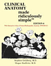
Each chapter is subdivided into Radiographic Anatomy, Approach and Specific Problem sections. In the Radiographic Anatomy sections, key anatomical landmarks are identified using simple illustrations. In the Approach sections, reading of the radiographic examination is explained using analogies, illustrative cartoons and mnemonics. In the Specific Problem sections, the radiographic findings of the most common and deadly conditions are discussed.
Topics such as MRI, CT, barium studies and ultrasonography are covered in greater detail with the companion Download of Clinical Radiology MRS Atlas program (Win/Mac).
Discover more
Ratings and reviews

- Flag inappropriate
About the author
Hugue Ouellette M.D.: Dept. of Diagnostic Radiology, Director of Education Musculoskeletal Radiology, Massachusetts General Hospital, Harvard Medical School.
Patrice Tetreault, MD, is Assistant Professor of Orthopedic Surgery, CHUM - Pavillon Notre-Dame, University of Montreal.